Electrophoresis of Nucleic Acids
Updated 11-07-2024 by: Rachel Bober
This protocol is based on Bio-rad.com photos instructions and procedures were taken from this link
General Comments
Unlike proteins, nucleic acids (being polyphosphoric acids) are all identically charged per mass unit. Therefore, their rate of movement in an electrical field is determined solely by mass (length), providing that they are all similarly folded. Since a ds structure is incapable (almost) of further folding, DNA is separated upon electrophoresis by size alone. 1% agarose has become quite standard for nucleic acid electrophoresis. Such electrophoresis can resolve fragments of 500 bp-10kb on a single gel. Nucleic acids absorb UV light at about 260nm, and consequently emit light at about 320 nm.
Buffer preparation
Prepare:

And dilute to 0.5XTBE buffer.
Procedure

- Use leveling bubble to a sure that the gel caster device and gel stage of gel running device are being leveled, otherwise adjust leveling feet and place device in a proper leveled position.
- In 250 ml Erlenmeyer flask, weigh 1 grams of agarose powder (for small size gel weigh 0.5 grams of agarose powder).
- Add 100 ml TBEx0.5 buffer and swirl to suspend the agarose powder in the buffer (for small size gel add 50 ml TBEx0.5 buffer). Caution: wear protective gloves, safety glasses and lab coat.
- Place flask with gel solution in the microwave. Use medium setting, set a timer to 2 minutes, stop the microwave oven every 30 sec to mix gently and to swirl the flask. This action is made to suspend the undissolved agarose thus gel solution should be transparent.
- Be aware that flask can be hot, and agarose was boiled, therefore hold the flask neck with a paper towel or with a cloth.
- Cool down gel solution to about 60°C by using a stream of tap water on the outer side of the flask (don’t inhale the vapors and don’t let tap water enter your gel solution!).
- Use 1-10 ul filter tips and 1-10 ul pipette to add 4ul abm’s SafeView™ Classic to large size gel solution and 2 ul for small size gel solution.
abm’s SafeView™ Classic - BioConsult, PO Box 7672, Jerusalem Israel 91076 Tel: 972-(0)2-5667043 Fax: 972-(0)2-5662790 Email: sales@bioconsult.co.il Website

- Swirl the flask gently to suspend the DNA Stains.

- Pour gel solution on the sealed UV-transparent gel tray. Avoid bubbles.
- Place a comb (it’s in the drawer under the gel camera).
- Wait 45 minutes until the gel solidify.

- Take out the comb.
- Place gel on the leveled gel stage of the running device.
- Fill both sides tanks of running device with ‘used TBEX0.5 buffer’.
- Submerge the gel in the UV-transparent tray (that placed on leveled gel stage) in buffer and make sure wells are fully covered with buffer. Remark: on this stage don’t add buffer up to maximum level, only after loading your sample.
- According to a writing plan, use filer tips and pipettor and load 2 ul of DNA ladder (marker) and 5-6 ul samples.
- Don’t forget to add sample buffer if needed. Usually the ratio is: 1 ul sample buffer to 5 ul of a sample = 6ul in total. Prepare drops on a parafilm, pipette for mixing and load into gel well.
- Cover gel with ‘used TBEX0.5 buffer’ up to maximum limit line.
- Place safety lid carefully without disturbing the gel. Make sure safety lid is place in the right orientation. Contact banana jacks on the safety lid to banana plugs in the base- black to black, red to red.
- On the side of the running device, search for an arrow sticker that points of the correct running direction. Pay attention to gel running direction and adjust it, if needed.
- Set voltage to 110 V and press run.
- Make sure that you see small size bubbles and that your samples are going out from the wells and runs properly in a straight line.
- When the front line reaches to 2/3 of the gel, after 60 minutes or more, switch power off or press on pause bottom.

- Take the gel in the UV-transparent tray out from the running device. Leve buffer in the running device.
- Transfer the gel out from the UV-transparent tray to a smooth transparent polypropylene plastic bag. Make sure the bag has no scratching and its UV-transparent. Avoid bubbles.
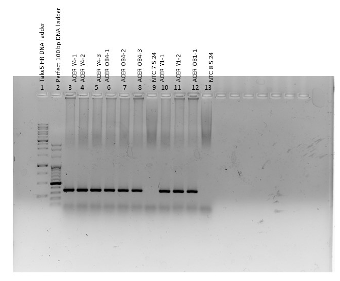
- Look for DNA bands under UV lamp.
- If gel band are observed, then use gel camera to record results.
Gel Camera
- Open camera drawer and place gel on the UV-transparent plastic bag. Use tray slits to find the proper position in the middle of the camera tray.
- Switch camera on.
- Gel camera has a touch screen
- Log in
- Let camera lamp warm up and then press continue.
- Select your folder.
- Select ‘safe red’ filter and automatic / manual exposure.
- Adjest photo parameters with cursor.
- Save photo on disk-on-key.
- Log out to main screen.
- While in main screen of the camera, switch off the camera.
Cleaning
- Dispose gel and clean camera tray.
- Collect ‘used TBEX0.5 buffer’ from running device to a bottle.
- Wash devices with distilled water to avoid salt remaining. While washing, be careful not to disconnect wires and electricity sockets.
- Use Kimwipes to dry equipment to avoid scratching.